IFMBE PROCEEDING, 2nd European Medical and Biological Engineering Conference, 2002, Vienna, P1544, 1545
__________________________________________________________________
ESTIMATION OF FETAL HEART RATE
USING ABDOMINAL ECG RECORDINGS
V.Kalakutskij, V.Konyukhov, E.Manelis
Samara State Aerospace University, Dept. of Radiotechnics, Samara, Russian Federation
Samara Regional Cardiology Centre, Dept. of Obstetrics, Samara, Russian Federation
Abstract:
method of an estimation of a fetus condition includes abdominal ECG registration, correlation processing of the received data, fetal R-R intervals allocation, estimation of distribution parameters and diagnostic index calculation, describing activity of sympathetic nervous system of fetus.Keywords: fetal ECG detection, abdominal ECG processing, fetal heart rate variability.
Introduction
Monitoring of a fetus condition by means of tracking the parameters of heart rate is considered to be one of the promising ways of modern antenatal diagnostics [1].
The suggested technique is based on definition of fetal heart rate variability (FHRV) and calculating indices, which have high diagnostic value. In this case maternal abdominal ECG signal could be used as a raw signal. Special signal processing algorithm is applied to obtain fetal ECG and measure RR-intervals of fetus.
The index of sympathetic department of vegetative nervous system regulation activity is calculated from the FHRV estimation and then is chosen to be the resulting diagnostic index.
Materials and Methods
The condition of an organism can be estimated with the help of the diagnostic parameter describing activity of vegetative regulation in physiological systems of an organism in reply to action of stress factors. One of the systems for an estimation of a fetal condition can be the cardiovascular system, process under control – heart rate, observed physiological parameter - time interval between two consecutive fetal heart beats (FHBI).
Duration of FHBI most precisely can be determined as an interval between successive R-peaks on fetal ECG. Chronotrope structure of fetal heart rate can be investigated with the help of studying of parameters of variability which occur due to various processes in multiplanimetric system of life processes regulation.
Methods of FHRV estimation in time domain turn to be easy in computing. In these methods sample of FHBI of the fixed volume is used as the input information.
There are well-known statistical approaches in formation of diagnostic parameters, which deal with transformation of FHBI sample into geometrical structure, such as the histogram, and definition of its parameters.
As the integral parameters of condition computed from the histogram data, most frequently are used [2]:
Hardware realization of the considered method includes fetal heart rate registration and the further processing of the received signal in real time using a specially developed algorithm.
Various methods of fetal ECG detection have been described. They include adaptive algorithms of maternal ECG processing together with correlation signal reception [3], another method based on application of subsequent singular value decompositions [4], and also application of wavelet transform [5].
It is necessary to emphasize that the primary goal of the above mentioned works was the detection of the full fetal ECG, including small and buried in myographic noise P, Q, S and T peaks. In our case it is enough to record only R-R intervals. The given condition facilitates a problem and makes possible algorithms simplification.
The technique developed consists of the following stages: choice of correct electrode placement on the abdominal surface, abdominal maternal ECG recording, blanking of maternal QRS-complexes, detection of fetal R-R of intervals.
The choice of points for ECG electrodes placement is a pretty complicated problem, which was investigated by many researchers. The early publications about the subject belong to Sureau and Troceillier (1958) which created a topographical map of abdominal surface and correlated amplitude of registered signals with the position of a fetus in a womb, position of mother during ECG registration and term of pregnancy. Development of algorithm for automatic search of fetal electric vector position instantly enables to determine electrode placement, which will give the maximal projection of fetal electric vector on the abdominal surface of mother.
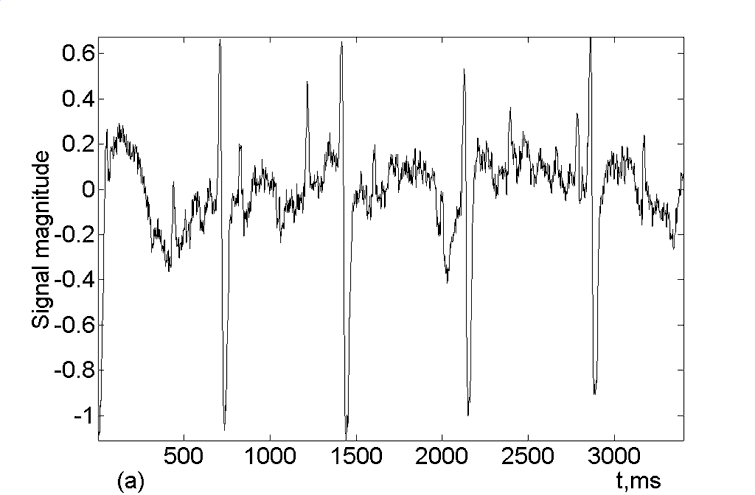
The device for the recording of composite ECG of mother and fetus consists of the low noise amplifier, 16-bit sigma-delta ADC and unit for data input into PC. The channel of registration has the following characteristics: sampling frequency 1000 Hz, adjustable factor of amplification from 1500 up to 48000, noise given to an input does not exceed 0.002 mV. The developed experimental device has a wide dynamic range that enables registration of fetal and mother ECG as well with a sufficient degree of accuracy (Figure 1a).
Blanking of maternal QRS-complexes (Figure 1b ) is conducted with the help of a correlation method, which gives position of maternal QRS-complexes on the time axis. The found interval is filled with points linearly interpolated between the edges of complex.
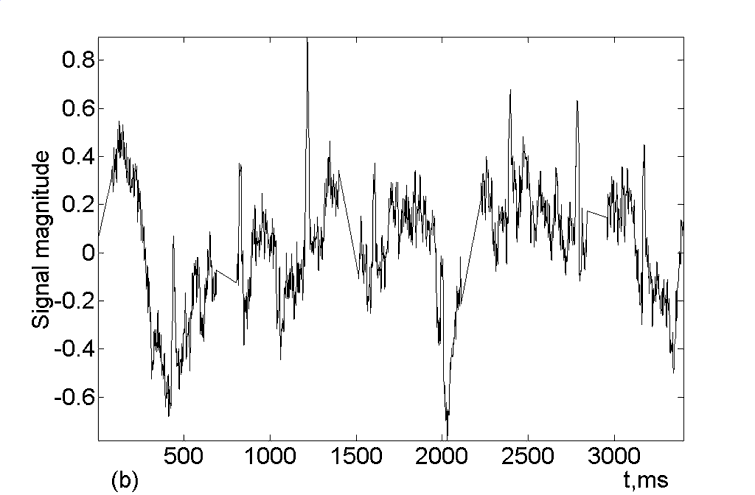
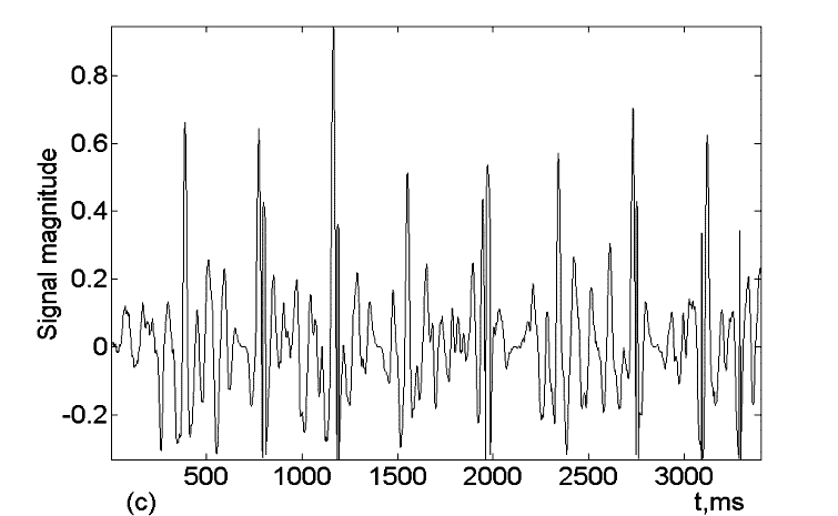
Figure 1: (a) the composite maternal abdominal ECG signal, (b) abdominal signal with blanked maternal QRS-complexes, (c) correlation function of maternal abdominal ECG signal with blanked QRS-complexes and sample of fetal QRS-complex.
The further definition of the fetal R-peaks on time axis is accomplished by finding of maxima of correlation function of fetal ECG and the averaged pattern of a fetal QRS-complex (Figure 1c).
The obtained FHBI values are used for formation of the histogram of FHBI distribution and the computing of SAI diagnostic parameter, serving as the indicator of activity of fetal sympathetic nervous system
Results
As a result of the carried out research 28 pregnant women with gestation terms from 26 to 41 weeks were surveyed.
In each case there was performed manual processing of FHBI as well as processing using the offered technique. While computing of diagnostic parameter SAI on the basis of the experimental data, the insignificant divergence in values was revealed which characterizes an error of a method.
Discussion
The technique of abdominal ECG processing was chosen for computing algorithm simplification and it takes into account the opportunity of real time mode realization with a minimum of computing operations. The clinical importance of antenatal FHRV estimation consists in the revealing of neonatal risk group, which is threatened by different kinds of pathological syndromes in perinatal period. Stable high parameters of sympathetic activity of a fetus can testify, for example, about growing oxygen insufficiency. Favorable change of the given parameters shows the improvement of fetus’s condition, as result of efficiency of the therapy treated.
Conclusions
The technique for an estimation of a fetus’s condition was applied. Abdominal ECG of a mother-to-be was selected as an input source, from which the fetal heart rate has been detected. The basic part of detection algorithm is the correlation analysis of the abdominal ECG and blanking of maternal QRS-complex. This technique is used in real-time mode and serves as an approach to the problem of fetal stress diagnostics by means of maternal abdominal ECG processing.
REFERENCES